Rosenberg X Ray View
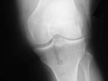
It should be the initial study for any patient with a suspicion of knee osteoarthritis.
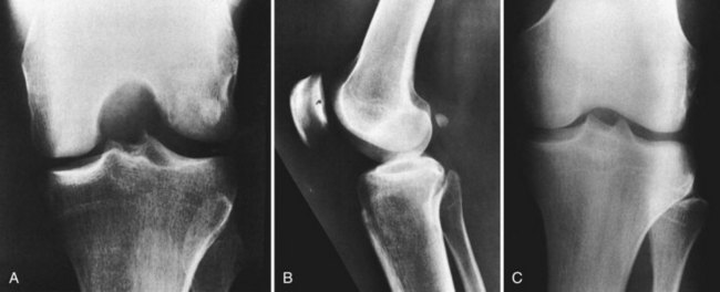
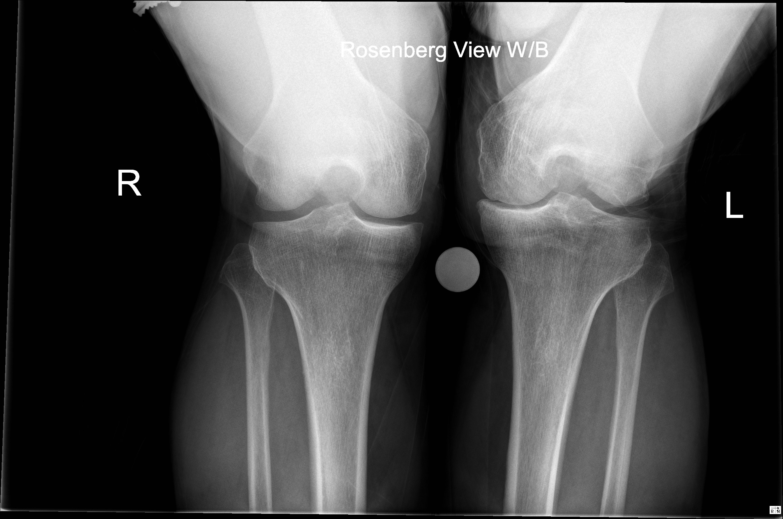
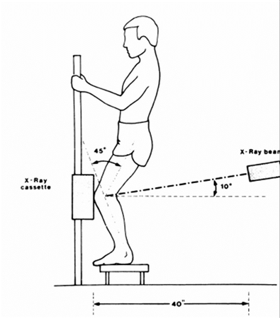
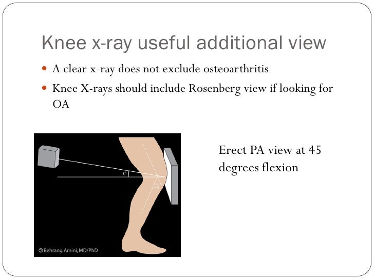
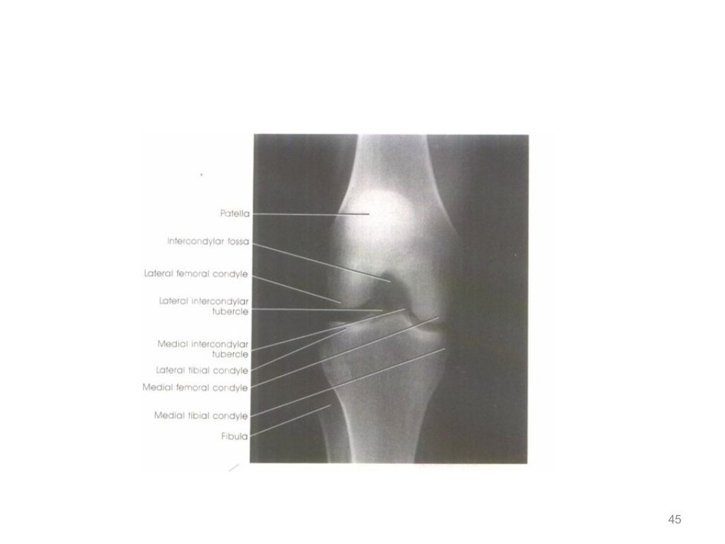
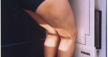
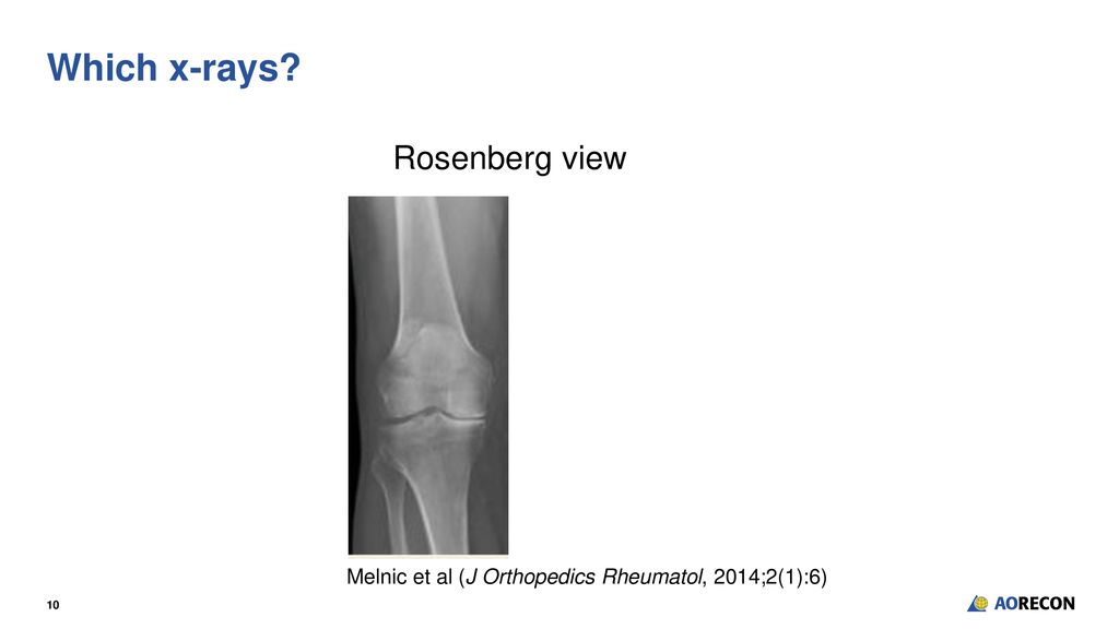
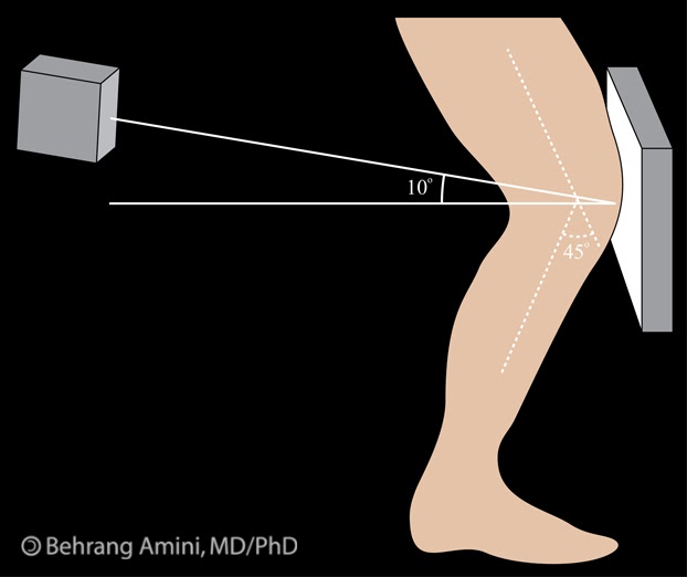
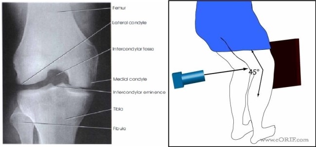
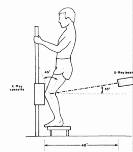
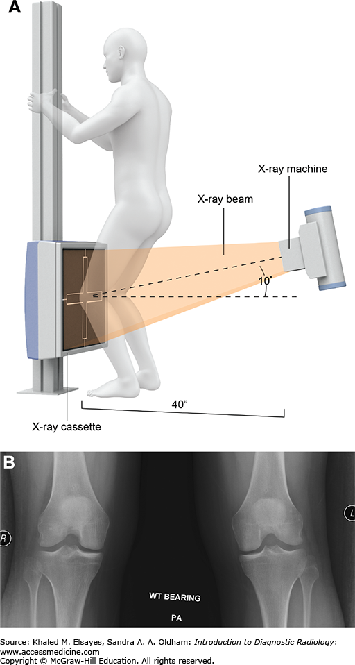
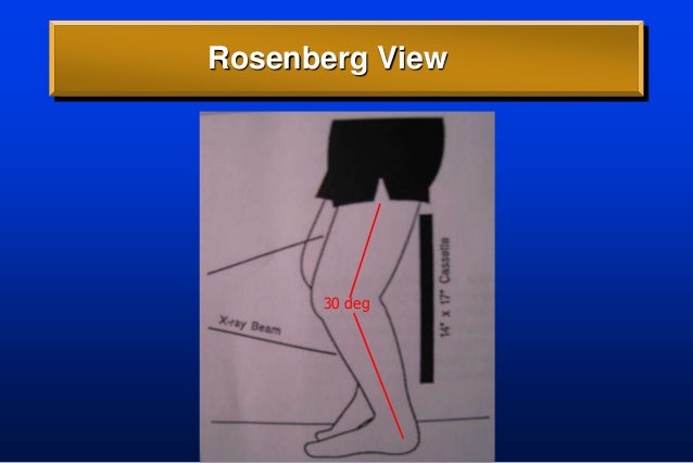
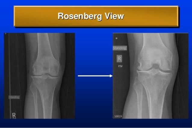
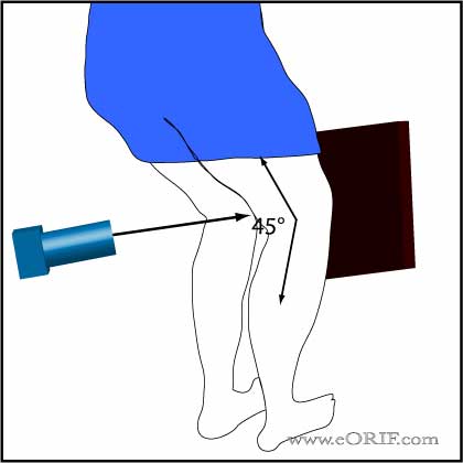
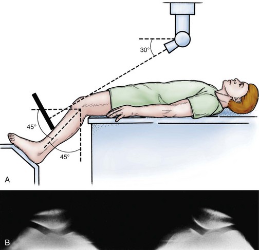
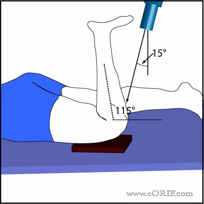
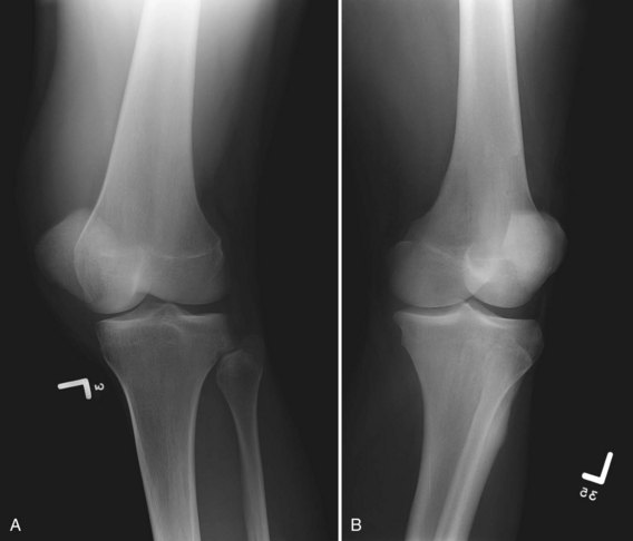
Rosenberg x ray view. The rosenberg view is a 45 degree flexion posteroanterior weight bearing view of the knee with the patellae touching the image receptor. All patients were symptomatic at the time of evaluation with a suspicion of knee osteoarthritis. Indications the rosenberg view is performed for. The patient stands with the knees bent to 45 degrees against the x ray plate while the x ray machine is behind the knee at an angle slightly downwards 450 flexion pa weight bearing view.
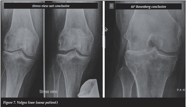
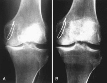
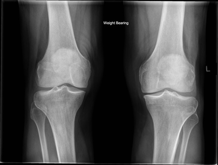
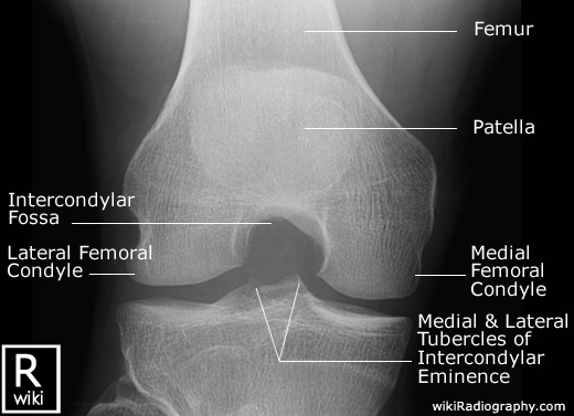
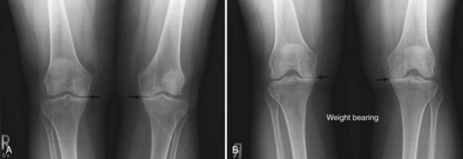
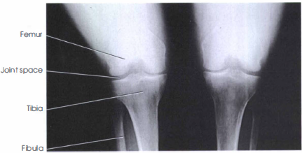
Advantages of the rosenberg view this x ray view may ofer information about narrowing of the normal joint space which is not available in a straight leg x ray. Pk view helps identifying the direction of femor and tibial tunnel as well as the depth and obliquity needed to obtain the best possible knee rotational stability. The pk view fig 7 shows the intercondylar notch and the correct direction and angle of the femoral tunnel certanly better than other x rays as the ap tunnel view or the rosenberg. Radiological classifications were according to ahlbaeck and kellgren lawrence.
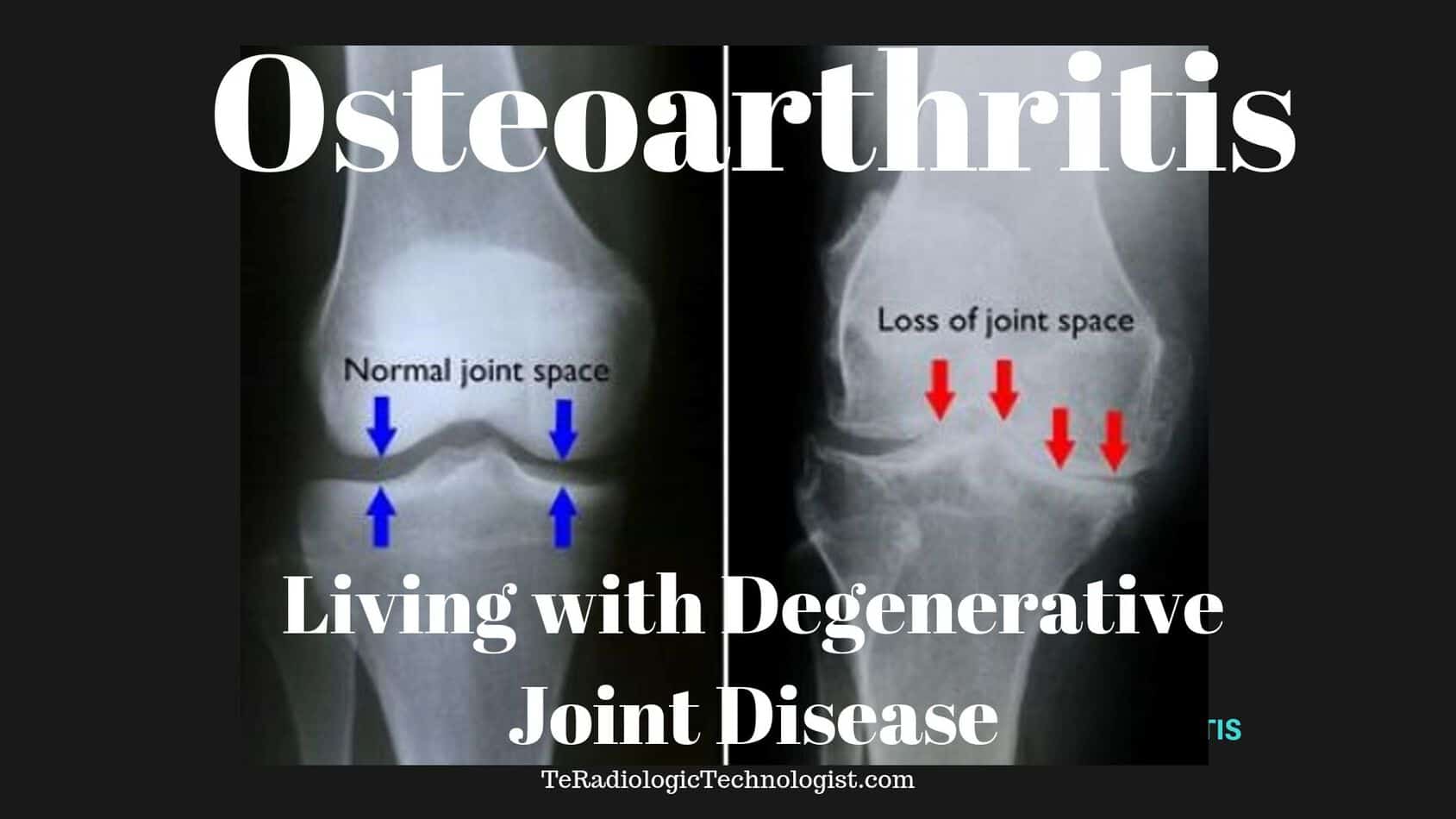
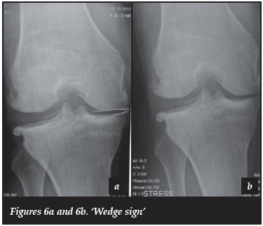
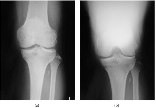
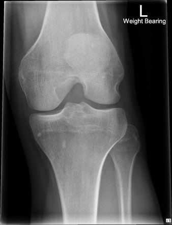
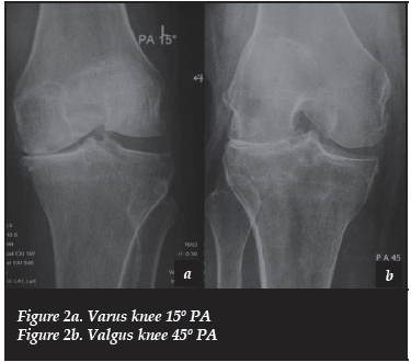
We evaluated 44 knees with conventional ap weight bearing in full extension and rosenberg pa weight bearing in 45 degrees of flexion x ray projections in 32 patients 24 women and 8 men aged 26 to 78 years. The rosenberg view of the knees is a specialized series often used to detect early signs of osteoarthritis. Weight bearing projection used to assess joint space related pathology such as osteoarthritis oblique view. The rosenberg view was created to address this issue.
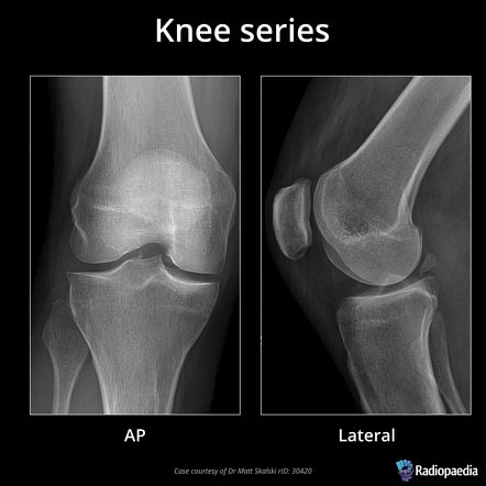
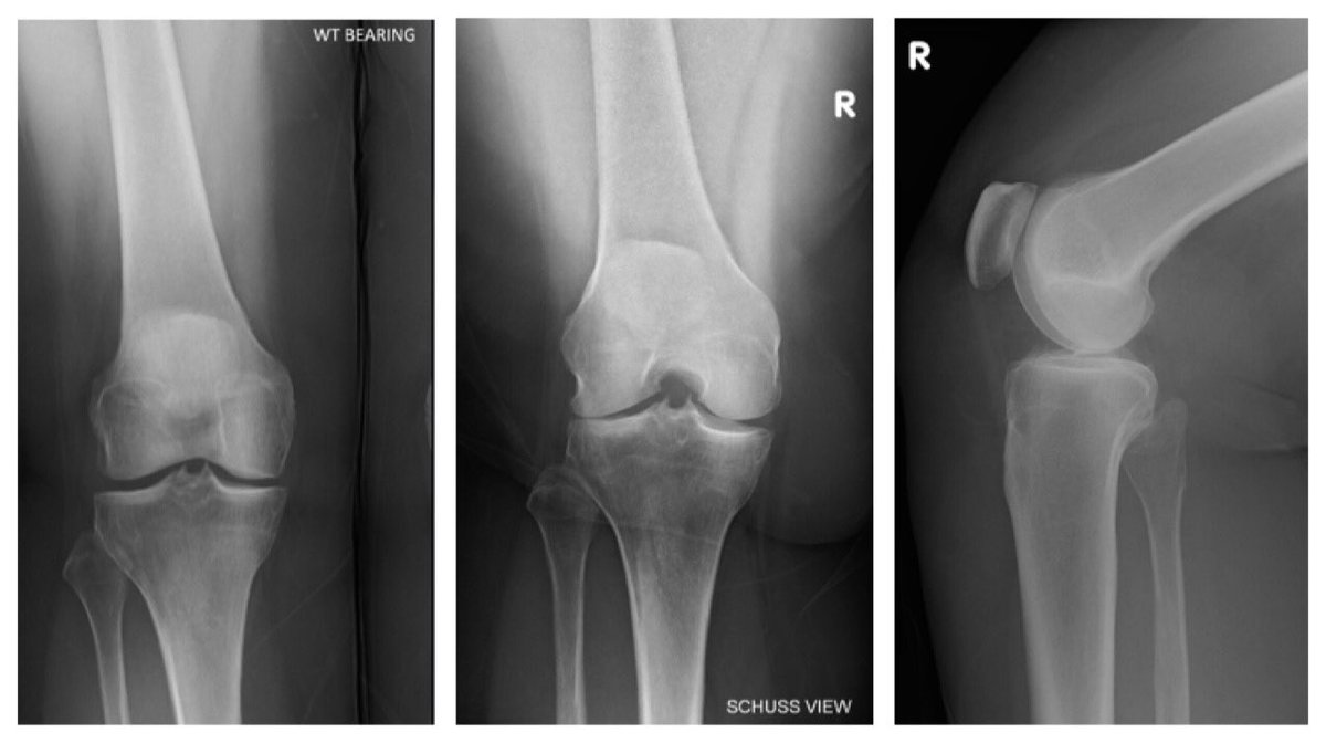
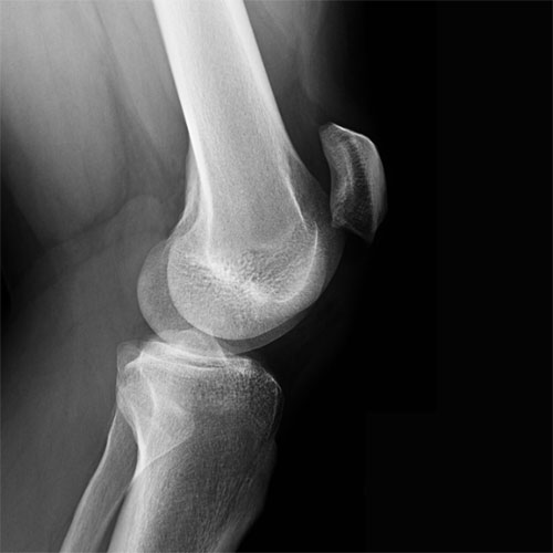
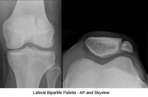
Radiological evaluation included ap weight bearing lateral knee rosenberg and sky view x rays. This 450 flexion posteroanterior weight bearing view of the knee is taken with the patella touching the image receptor. View utilized to demonstrate intercondylar space often used for oa and suspected tibial plateau fractures rosenbergs view. The x ray tube is 40 inches 1016 cm away from the image receptor centered at the patellae and pointing caudad 10 degrees.
The x ray tube is 40 inches 1016 cm away from the image receptor which is centered at the patellae and pointing 100 caudad. The rosenberg view is a 45 degree flexion posteroanterior weight bearing view of the knee with the patellae touching the image receptor.

